| microscope base |
| bottom of the microscope |
| microscope arm |
| with the base, used to carry the microscope |
| microscope stage |
| holds the slide |
| microscope body tube |
| transmit the magnified image |
| microscope condenser |
| lenses that focus light into a cone |
| microscope iris diaphragm |
| controls the angle and size of the cone of light |
| microscope revolving nosepiece |
| holds objective lenses |
| microscope obective lenses |
| magnify & invert the image |
| microscope focal point |
| formed when light rays converge at one point |
| microscope coarse adjustment |
| larger knob used to focus on low power |
| microscope fine adjustment |
| smaller knob used to focus with high power and oil immersion |
| microscope field of vision |
| area seen through the microscope |
| microscope magnification |
| the number of times an image is increased in size |
| formula for magnification |
| determined by multiplying the power of the objective by the power of the ocular lens |
| resolution |
| ability to distinguish two points as distinct and separate |
| refractive index |
| the amount light bends when it enters a new medium |
| parfocal |
| when one lens is focused, all the other lenses will also be in focus |
| which lens has the shortest focal distance? |
| oil immersion |
| the three basic bacterial shapes |
| rods (bacilli), spheres (cocci), spirals |
| the field of vision decreases when the magnification... |
| increases |
| why does immersion oil increase resolution? |
| it has the same refractive index as glass (1.52) and the light does not bend between the slide and the objective lens |
| when viewing large organisms like fungi or protozoa, it is best to use the ___________ lens |
| low power |
| spherical aberration |
| when the middle of the field of view is in focus but the periphery is blurry. Light passing through the middle of the lens has a different focal point than light passing through the outside |
| chromatic aberration |
| many colors appear in the field. occurs when each wavelength of light has a different focal point |
| In bright field microscopy, the image is made from: |
| light that is transmitted through a specimen |
| Bacterial stains will _____ the organism |
| kill |
| The condenser lens ___________ the light |
| concentrates |
| Refraction is ________ of light rays |
| bending |
|
A microscope produces 2 images. One is _____ and one is ______. |
|
real virtual |
| The virtual image appears ___________ the microscope |
| below or within |
| The formula for calculating magnification: |
|
total magnification =
magnification by the objective lens x magnification by the ocular lens |
| Resolution is defined as: |
| clarity of an image |
| The limit of resolution is: |
| an actual measurement of how far apart two points must be for the microscope to view them as being separate |
| Write the formula for the limit of resolution: |
|
λ D=----------------------------------- NAcondenser +NAobjective |
| Numerical Aperture is: |
| a measure of a lens's ability to "capture" light coming from the specimin and use it to make the image |
| Using immersion oil makes the numerical aperture__________ |
| increase |
|
In dark-field microscopy, objects appear ________ against a _________ background |
|
brightly lit dark |
| In phase contrast microscopy, the specimen appears as various levels of ______ against a bright background |
| "darks" |
|
Fluorescent microscopy uses fluorescent ______ that emit light when illuminated with _____________ light |
|
dye ultraviolet
|
| A mixed culture contains: |
| two or more species |
| A pure culture contains: |
| only a single species |
| The purpose of streaking bacteria on a plate is to: |
| isolate an individual species from a mixed sample |
| Individual cells grow into: |
| colonies |
| CFU stands for: |
| colony-forming unit |
| A CFU consists of: |
| individual cells or pairs, chains, or clusters of cells |
| Ubiquitous: |
| Organism can be found everywhere, could be isolated from soil, water, plants, and animals |
| Define pathogenic: |
| capable of causing disease |
| Define opportunistic pathogen |
| capable of causing disease if introduced into a suitable part of the body |
| define reservoir: |
| any area where a microbe resides and serves as a potential source of infection |
| Pellicle |
| organisms float on top and produce a surface membrane |
| sediment |
| organisms sink to bottom |
| turbidity |
| evenly distributed throughout |
| flocculent |
| suspended chunks |
| Organisms that can infect us: |
|
Amoeba (Entamoeba histolytica causes dysnetery) Nematodes (Enterobius vermicularis - pinworm - intestines Ascaris lumbricoides - intestines Necator americanus - intestines Trichinella spirallis - muscles) Ciliates (Balantidium coli - intestines)
|
| Organisms that may transmit disease |
| arthropods |
| most commonly used staining method |
| gram staining |
| gram staining - which stain is applied first? |
| crystal violet |
|
gram staining
what forms inside the cell after you add iodine?
|
| crystal violet-iodine complex |
|
gram staining
what type of cell is decolorized? |
| gram negative |
|
gram staining
Name the counterstain |
| safranin |
| what effect does alcohol have on the gram-negative cell wall? |
| the alcohol extracts the lipid, making the gram negative cell wall more porus and unable to retain the crystal-iodine complex, decolorizing it |
| Explain why gram-positive cells are not decolorized |
| the thicker peptidoglycan traps the crystal violet-iodine complex more effectively, making them less susceptible to decolorization |
| What color will gram-positive cells be if the decolorizer is left on too long? |
| reddish |
| Describe the appearance of a good emulsion |
| dries to a faint haze on the slide |
| what happens to older gram-positive cultures? |
| may decolorize and give a gram negative result |
| In the negative staining technique a chromogen (dye) has a ____________ charge. |
| negative |
| The pH of negative stains is_____________ |
| acidic |
| Negative stains do not enter bacterial cells because the charges ____________ each other. |
| repel |
| Negative staining is commonly employed for bacteria that are: |
| too delicate to withstand heat-fixing |
| Acid-fast bacteria have ____________ in their cell walls |
| mycolic acid |
| Acid-fast organisms resist _________ by _________ alcohol. |
| decolorization, acid |
| The names of the 2 acid-fast staining procedures are: |
|
Ziehl-Neelsen (ZN) Kinyoun (K) |
| When preparing an acid-fast smear, a drop of __________ is used to help the ____________ organisms adhere to the slide |
| serum, slippery |
| The primary stain in the ZN method is _________ because it is soluble in _____________ |
| carbolfuchsin, lipid |
| Heating causes acid-fast cell walls to _________ |
| melt |
| the counterstain in an acid-fast stain is |
| methylene blue |
| Acid fast cells are colored |
| reddish purple |
| Non acid-fast cells are |
| blue |
| Capsules are made of __________ or _________ |
| mucoid polysaccharides, polypeptides |
| (Capsule stain) Two examples of netgaive stains are: |
| Congo red, nigrosin |
| (Capsule stain) Negative stain pH is ___________ and they stain the background |
| acidic |
| (Capsule stain) A basic stain is used to stain _________ |
| the cell |
| (Capsule stain) We do not heat fix because: |
| it causes the cells to shrink, leaving an artifactual white halo that may be interpreted as a capsure |
| Cells stick to the slide by adding a drop of ___________ |
| serum |
| An endospore is: |
| A dormant form of the bacteria that allows it to survive poor environmental conditions |
| Endospores are covered with a protein called: |
| keratin |
| (endospore stain) The primary stain is called: |
| malachite green |
| (endospore stain) The decolorizer is: |
| water |
| (endospore stain) The cells that are counterstained with safranin are ______________ and _____________ |
| vegetative cells, spore mother cells |
| Location of endospore: central |
| In the middle of the cell |
| Location of endospore: terminal |
| at the end of the cell |
| location of endospore: subterminal |
| between the end and the middle |
| Two spore shapres are: |
| spherical, elliptical (oval) |
| some spores are large and make the cell look: |
| swollen |
| Why can't we view flagella using an unstained preperation? |
| flagella are too thin to be observed with light microscope and ordinary stains |
| flagella - monotrichous |
| one flagellum at one end |
| flagella - amphitrichous |
| flagella at both ends |
| lophotrichous |
| tufts of flagella at one end |
| peritrichous |
| flagella all over the cell |
| Why does light of a shorter wavelength produce a clearer image than light of longer wavelengths? |
| As wavelength gets smaller, resolution gets smaller because wavelength is on the top of the equation |
| Colony morphology includes: |
| colony size, color, shape, margin, elevation, texture |
| colony morphology - shape |
| round, irregular, punctiform |
| colony morphology - margin |
| entire, undulate, lobate, filmentous, rhizoid |
| colony morphology - elevation |
| flat, raised, convex, pulvinate (very convex), umbonate (raised in center) |
| colony morphology - texture |
| moist, mucoid, dry |
| colony morphology - color |
| opaque, translucent, shiny, dull |
| colony morphology - other factors |
| length of incubation, temperature of incubation, type of medium grown on, oxygen concentration during incubation |
| Why are microorganisms located on the desks not sterilized as extremely as the plates? |
| Bugs that grow on desks at 25 degree C are probably not human pathogens. Plates have many more bugs on them as well. |
| What is significant about organisms that grow well at 37 degrees C? |
| They probably came from humans. |
| Capsule stain - why must the sample be emulsified in serum? |
| To help them stick to the slide because they are slippery. |
| Why do oral bacteria produce a capsule? |
| protection against phagocytocis and to stick to surfaces and each other forming a biofilm |
| Why was an older culture of Bacillus used to demonstrate endospores? |
| Spores are formed in response to nutrient depletion, so the |
| Why can't flagella be observed in action? |
| Because they are too thin to be seen with regular stain. A mordant must be used to encrust the flagella so it is thick enough to be seen. |
|
Type of microscopy:
|

| bright field microscopy |
|
Type of microscopy:
|

| dark field microscopy |
|
type of microscopy:
|

| fluorescence microscopy |
|
type of microscopy:
|
| phase contrast microscopy |
|
bacterial morphology:
|

| gram positive cocci |
|
bacterial morphology
|

| ovoid coccus (Lactococcus lactis) |
|
bacterial morphology
|

| gram positive bacilli (Bacillus) |
|
bacterial morphology
|

| gram positive staphylococci |
|
bacterial morphology:
|

| gram positive streptobaccillus |
|
bacterial morphology:
|

| gram positive spirilla |
|
bacterial morphology: |
| spirochetes |
|
bacterial morphology:
|

| gram negative vibrio (Vibrio cholera) |
|
bacterial morphology:
|

| gram negative diplococci (Nesseria gonorrhea) |
|
bacterial morphology:
|
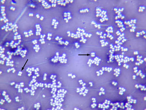
| tetrads (Micrococcus roseus) |
|
bacterial morphology:
|
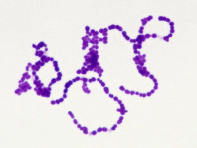
|
gram positive streptococci (Streptococcus pyogenes) |
|
bacterial morphology
|

|
gram positive bacilli, palisades arrangement (Corynebacterium) |
| How to do a plate streak: |
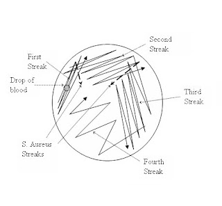
|
|
Broth growth:
|

|
1- obligate aerobes (need oxygen) - growth at top 2 - faculative anaerobes - growth throughout, but more growth at top 3- microaerophiles 4 - anaerobes - growth at bottom, no growth at top where oxygen is present |
| Gram stain procedure |
|
1 - heat fix emulsion 2 - cover smear with crystal violet stain for 30-60 sec 3 - rinse with distilled water 4 - cover smear with iodine for 30 - 60 sec 5 - rinse with distilled water 6 - decolorize with alcohol 7 - counterstain with safranin for 30 - 60 sec 8 - rinse with distilled water 9 - blot dry with bibulous paper |
|
gram positive vs gram negative results:
|

|
gram positive - dark purple gram negative - pinkish red |
|
Negative stain:
|

| Bacteria are unstained against dark background |
|
acid-fast stain (ZN)
|

| in ZN stain, acid fast cells are reddish-purple (non acid fast cells are blue) |
|
acid fast stain (K)
|
| acid fast cells are reddish purple (non acid fast cells are blue) |
|
capsule stain:
|

| acidic stain colorizes the background while the basic stain colorizes the cell, leaving the capsules as unstained white clearings around the cell |
|
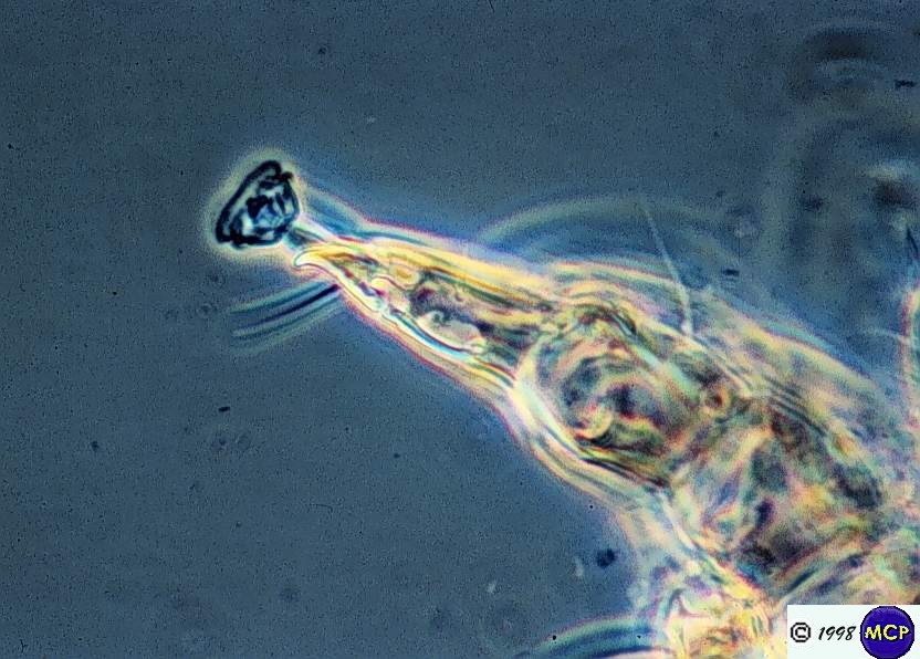
Flagella stain:
|

| peritrichous flagella |
|
flagella:
|

| monotrichous |
|
flagella: |
| amphitrichous |
|
flagella:
|

| lophotrichous |
|
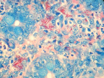
Endospores:
|

| terminal swollen |
|
endospores
|

| central |